
ErlanggaBlog.com – There are many symptoms that are related to Superficial Infrapatellar Bur-sitis. Some of these symptoms can include limited range of motion or muscle weakness, and you should see a doctor if you experience these symptoms. A physician will prescribe an appropriate treatment that can alleviate pain and inflammation in the joint. Depending on the severity of your condition, you may require a variety of treatments.
Symptoms associated with superficial infpatellar bursitis
Acute or chronic trauma can cause the swelling of the infrapatellar bursa. A chronic hemorrhagic case may mimic a soft tissue sarcoma. There are several types of symptoms associated with superficial infrapatellar bursitis, including pain while kneeling and limited range of motion. Symptoms of this condition often co-exist with other conditions, including arthritis and tendinitis.
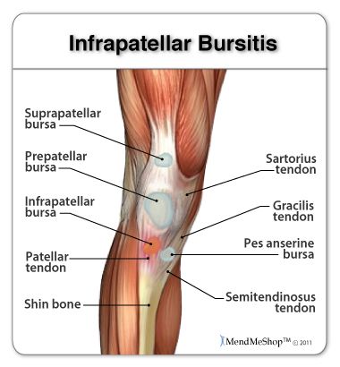
The most common symptoms of Superficial Infrapatellar Bur-sitis involve knee pain and swelling. This inflammatory condition affects the patellar tendon, which is in the outer part of the knee. The deep infrapatellar bursa, which surrounds the patellar tendon, may also be involved. If the inflammation is persistent, it may lead to calcification.
In some cases, a fibrous sheath may form and surgical removal is necessary
Initial treatment for Superficial Infrapatellar Bur-sitis is injection of corticosteroids to reduce the inflammation. In some cases, a fibrous sheath may form and surgical removal is necessary. After the initial treatment, the patient may need repeated aspirations. The goal is to reduce the inflammation and restore function. If you have persistent pain, your doctor may recommend surgical drainage and removal of the bursa.
Ultrasound imaging may be used to confirm diagnosis. Ultrasound imaging of the patellar tendon can help the physician better understand the extent of the inflammation and identify the cause. Ultrasounds may also be used to detect soft-tissue foreign bodies in the knee. The sonography results may also help detect the underlying abnormality. Once you’ve determined the type of bursitis, ultrasound guided movement may be helpful.
Percutaneous guided treatments have also been used for pain relief
Ultrasound-guided aspiration is an option for patients with symptoms of Superficial Infrapatellar Bur-sitis. Ultrasound-guided aspiration, may be used to remove the fluid and relieve symptoms. Local injection of long-acting analgesics and steroids may also be helpful. Percutaneous-guided treatments have also been used to relieve pain and eliminate the need for surgery.
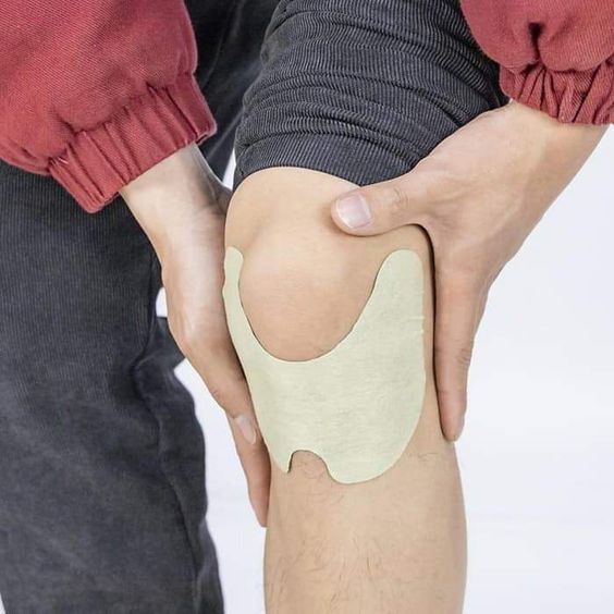
Cold therapy may help reduce pain and swelling. Improving blood flow to the affected area can improve your recovery time and improve soft-tissue health. Proper blood flow can also minimize the development of scar tissue and prevent atrophy of the tissue. A T*Shellz Wrap can also be helpful in relieving pain and inflammation and reducing the risk of re-injury. So, cold packs can reduce swelling and promote healing.
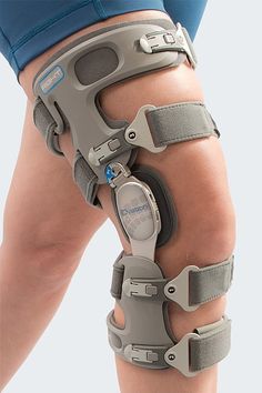
Non-surgical treatment includes rest
Treatment for Superficial Infrapatellar Bur-sitis varies depending on the severity of the symptoms and the cause of the pain. Usually, conservative management is successful for patients whose symptoms develop gradually and over a 24-48-hour period. Non-surgical treatments include rest, ice, and NSAIDs. Corticosteroid injections may be necessary when conservative measures fail. Besides rest and physical therapy, training modifications may also be beneficial.
A physical examination for Superficial Infrapatellar Bur-sitis includes a number of diagnostic tests. Your physician will likely perform an Ober test to assess the extent of ITB tightness. During this test, you will be asked to move your knee with either an arm or a leg, abducting or extending the knee. Once the test is complete, you will be asked to relax your knee and let the affected leg fall passively or with gravity. If your doctor suspects a discoid meniscal tear, you will be able to perform a McMurray test. The radiograph may show abnormalities including calcification of the patellar ligament and elevation of the tibial tubercle.


